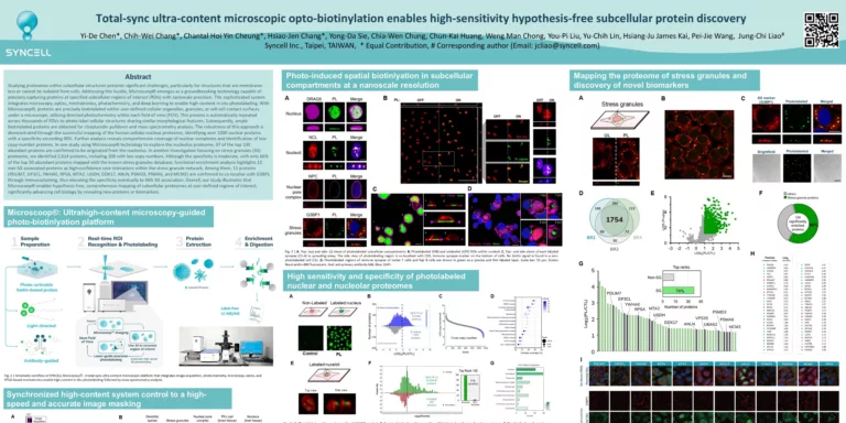
【Method】Total-sync ultra-content microscopic opto-biotinylation enables high-sensitivity hypothesis-free subcellular protein discovery
High-sensitivity, hypothesis-free spatial proteomics by integrating microscopy-guided photo-biotinylation with LC-MS/MS analysis identifies and maps proteins within compartments such as nuclei, nucleoli, stress granules, and primary cilia.
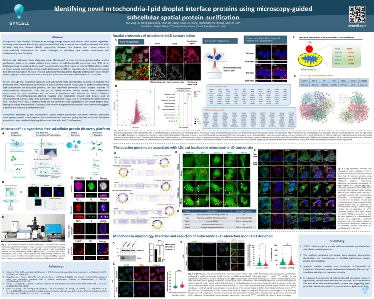
【Liver】Identifying novel mitochondria-lipid droplet interface proteins using microscopy-guided subcellular spatial protein purification
Integrating real-time image segmentation, two-photon photolabeling, and mass spectrometry-based proteomics, Microscoop reveals key regulators of mitochondrial-LD interactions, providing new insights into lipid metabolism and inter-organelle communication.
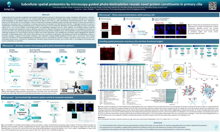
【Cilia】Subcellular spatial proteomics by microscopy-guided photo-biotinlaytion reveals novel protein constituents in primary cilia
Microscoop® selectively labels and analyzes primary cilia proteins within subcellular regions of interest, uncovering new ciliary proteins and their interactions.
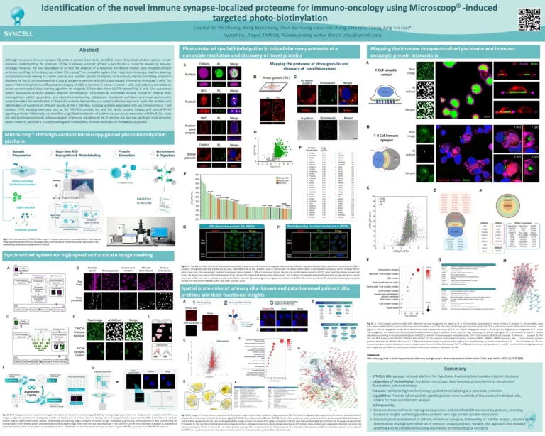
【immune synapse】Identification of the novel immune synapse-locatiized proteome for immuno-oncology using Microscoop-induced targeted photo-biotinylation
A new study shows how Microscoop® maps the poorly understood immune synapse-localized proteome more with nanoscale precision, discovering previously uncharacterized proteins and protein-protein interactions, offering new insights into immune synapse biology and potential therapeutic targets in immune-oncology.
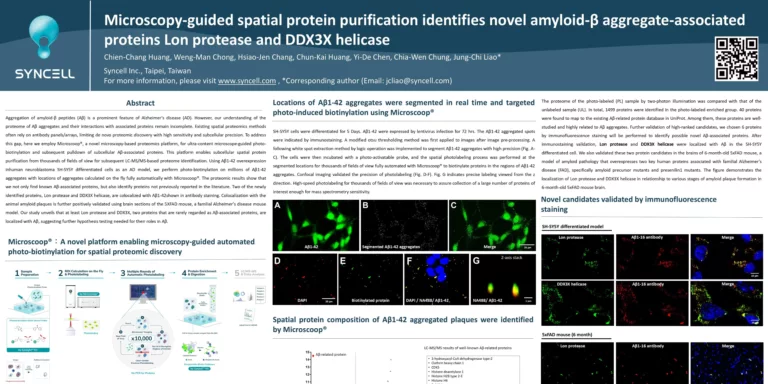
【Abeta】Microscopy-guided spatial protein purification identifies novel amyloid-β aggregate-associated proteins Lon protease and DDX3X helicase
Microscoop® enables microscopy-guided automated photo-biotinylation to identify proteins associated with amyloid-β (Aβ) aggregates, a hallmark of Alzheimer’s disease, including Lon protease and DDX3X helicase using AI-driven segmentation and high-precision mass spectrometry.