
The intricate network of proteins within the human body plays a critical role in maintaining health. Disruptions in this system can lead to the development of various diseases, particularly when misfolded proteins disrupt cellular functions. The accumulation of these misfolded protein aggregates is a hallmark of neurodegenerative diseases such as Alzheimer's, Parkinson's, amyotrophic lateral sclerosis, and Huntington's disease. These aggregates are known to contribute to the formation of toxic cellular environment, ultimately driving disease progression. Diseases characterized by protein aggregates pose significant challenges in biomedical research. For instance, in Alzheimer's disease, β-amyloid plaques and tau tangles accumulate within the brain, disrupting neuronal communication and leading to cognitive decline. Similarly, in Parkinson's disease, α-synuclein aggregates known as Lewy bodies develop within neurons, contributing to the motor symptoms and neurodegeneration characteristic of the disease. Cardiac amyloidosis arises from the accumulation of misfolded proteins in the heart, impairing its ability to pump effectively, which can lead to heart failure and death. From the neurodegenerative Alzheimer's to the insidious growth of protein deposits in heart failure, understanding the specific proteins involved in these aggregates is a fundamental step towards developing effective treatments.
Spatial Proteomic Discovery: Precision and Localization in Protein Identification
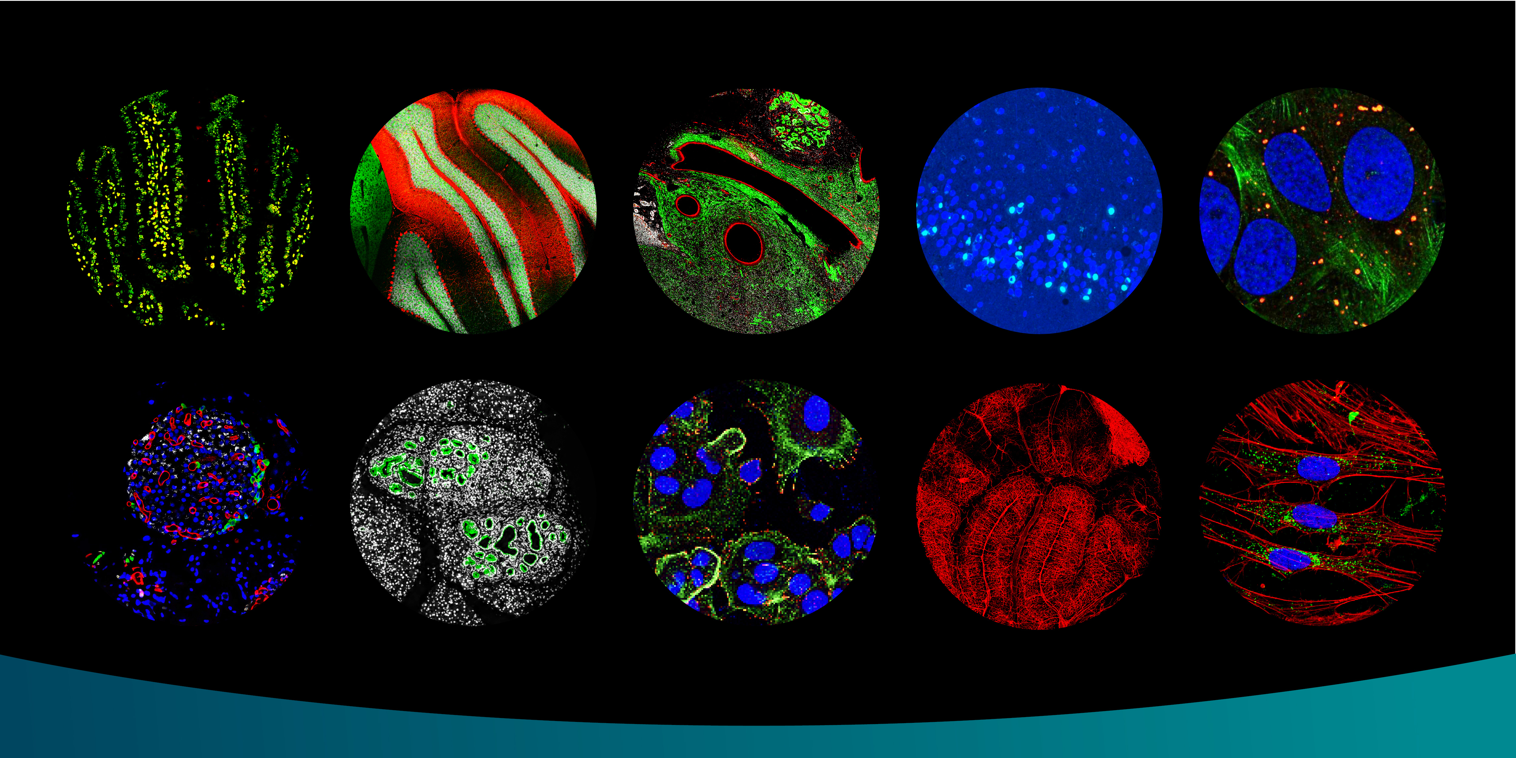
Spatial proteomic discovery of protein constituents in disease models represents a promising approach to tackle these complex conditions. This technique enables researchers to unravel the "what" and "where" of proteins fundamental to disease pathology. By leveraging advanced image processing, researchers can precisely identify region of interest, such as the specific protein aggregates. These regions are then targeted for precise photo-biotinylation, a process that utilizes light to activate a specialized photo-activatable probe that tags biotin to the proteins within the targeted region. Once the proteins are tagged with biotin, the biotinylated proteins can be pulled down using streptavidin beads and identified via mass spectrometry.
This pinpoint accuracy is not just a matter of resolution but a directed intention to understand the spatial distribution of proteins. The ability to conduct precise, localized photo-biotinylation and subsequent protein identification allows researchers to obtain detailed maps of protein interactions and distributions within affected tissues. This level of detail is essential for identifying key protein constituents involved in disease mechanisms and for developing targeted therapeutic strategies.
Conclusion: Advancing Biomedical Research
Spatial proteomic discovery of protein constituents in disease models represents a significant leap forward in biomedical research. This approach offers unprecedented precision and insights into the spatial organization of proteins within cells and tissues. As we delve deeper into spatial proteomics, we move closer to unraveling the complexities of various diseases and developing targeted treatments. By bridging the gap between molecular biology and clinical applications, spatial proteomics offers new dimensions for therapeutic intervention. Detailed mapping of protein interactions and distributions within diseased tissues can significantly accelerate our understanding of disease mechanisms.
| Info@syncell.com | |
| 617-631-2746 | |
|
|
200 Dexter Ave, Watertown, MA 02472, USA |
|
|
|